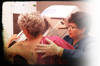
Should I worry about a mammogram call back?
What happens after either the doctor or I find a breast lump?
There are numerous ways your case can proceed. This is an important decision point. You must prepare yourself to make a decision about how you want to proceed if, after the examination, the doctor suggests there is a possibility that the lump may be cancer. You need to decide, for example, whether you wish to have a second opinion on the kind of treatment you will have.Is a second opinion important in breast cancer?
In our view, a second opinion is always important. It is more important in treating breast cancer simply because there are many options and differences of opinion in this area. A second opinion can help you think through your choices.What will the doctor do if he has any doubts about my lump?
If the doctor has any doubts, he will suggest further studies. These may include a mammogram, aspiration of the lump with a needle and syringe to see if it is a cyst, or an excisional biopsy. These tests are used to make sure that the lump is not cancerous or to determine treatment if it should be cancerous. Remind yourself again that eight times out of ten it usually turns out that the lump is not malignant.I am still confused. Tell me again what the steps are that would lead up to the operation for breast cancer. Let us take it from the time when you find a lump or something unusual about your breast and go to the doctor.
He will probably proceed as follows:
• He will look at what you have found and examine both breasts for lumpy areas.
• If he has any doubts, he may advise further studies, such as mammography, aspiration of the lump with needle and syringe to see if it is a cyst, a needle biopsy, or an excisional biopsy (taking out the whole lump for examination).
• If the biopsy shows that the lump is cancerous, the doctor will order a bone scan, x-ray of the chest, blood studies, and other tests to determine whether or not the cancer has already spread to distant parts of the body such as the bone, lungs, and liver.
What is a mammogram?
A mammogram is a soft tissue x-ray of the breast. It shows breast masses and helps to identify those which may be malignant. Mammograms can show tiny concentrations of calcification or perhaps other abnormalities that may indicate a tumor in the breast. Tumors can often be observed by this x-ray technique before they can be discovered by physical examination.Can a mammogram tell whether or not cancer is present?
Mammograms are diagnostic tools. They may indicate to a trained doctor whether cancer is present or not. They are used by surgeons to locate the site of the tumor and to check if there are additional tumors in the breast. However, they should never be used alone. They should be used in addition to a careful breast examination by a doctor who regularly treats breast cancer. In order to make a definitive diagnosis of breast cancer, a biopsy must be done of those suspicious areas seen on the mammogram.What is a Xeroradiograph?
A Xeroradiograph is a mammogram which basically uses the Xerox techniques. It processes the x-ray image on Xerox paper. A selenium coated metal surface is substituted for x-ray film, and after being exposed to x-rays, it is dusted with calcium carbonate powder. This produces an etchinglike image. The Xeroradiograph is blue and white. The mammogram is black and white. Sometimes this test is called Xeromammography.Who performs mammography?
Radiological technologists who are specially trained in mammography normally conduct the tests and process the mammograms. The mammograms are then given to a radiologist, who studies them, interprets them, and reports the findings to your own physician. It is very important that you go to a qualified radiologist who reads many mammograms, for as with other diagnostic tools, the results are only as good as the skill of the person who conducts and interprets the tests.What is digital mammography?
Digital mammography uses sensitive detectors rather than film or a Xerox type device to measure x-rays passed through the breast. It is a new, developing technique which should increase the amount of information while reducing the radiation received.Can mistakes be made in reading a mammogram?
As with any diagnostic tool, errors can be made in mammography or Xeroradiography. For instance, the position of the breast on the plate can distort results. The doctor reading the mammogram can make a mistake. This is why both the technicians involved in taking the films and the radiologist who reads them must be extremely qualified. Interpreting the films requires skilled and trained persons who read mammograms daily and can see the subtle differences.Your first mammogram: What to expect
Why is it a good idea to have a mammogram before having the breast lump removed?
A mammogram of the breast before surgery can serve several purposes:
• It can be used as a record for the doctor to use in future comparisons.
• It can tell if there are additional lumps in the areas of the breast which cannot be felt.
• Used in conjunction with other findings, it can sometimes help to determine if the lump is benign or cancerous.
• It will guide the surgeon during the operation.
What does the doctor look for in the mammogram to help him determine if the lump might be cancerous?
The doctor knows that benign tumors tend to show sharp edges in x-rays and are frequently surrounded by a halo of fat and seem to be homogeneous in density. Malignant tumors usually look as though they have tentaclelike tissue reaching into the surrounding areas. Fine, sandlike calcium deposits can be seen, and the skin in the area is often distorted. Even with his expert eyes, however, the doctor needs other guidelines besides the x-ray to help him make a positive diagnosis.What dose of radiation does a woman get when she has a mammogram done?
The dose of radiation for mammograms should be less than 1 rad per breast, with two views of each breast on a machine dedicated to mammography. You should ask when your physician makes the appointment for your mammogram or you should call the radiologist yourself before you have the examination to be certain that the machine being used is used only for mammograms. If the dose is more than 1 rad per examination, try to find another facility with lower dose equipment. Many "dedicated" machines today give considerably less than 1 rad per two view examination.What does the mammogram tell the doctor that he can't learn in some other way?
If the doctor finds a suspicious lump, the mammogram may give the doctor some indication as to whether or not the lump is cancerous. He will also see whether or not there are other lumps which might be cancerous in the breast and which cannot be felt by manual examination. This information is important to the doctor if he is to perform an operation on the breast. If the woman has large breasts or fibrocystic disease, mammography is especially useful.Can the doctor tell definitely from a mammogram whether or not cancer is present?
A negative mammogram does not guarantee there is no cancer in the breast. A mammogram can identify certain lumps as benign. However, the mammogram is only one phase in the total picture. In most cases, surgical biopsy is necessary to finally determine whether or not a suspicious lump is cancer.I have heard so much controversy over mammography that I don't know whether or not to have a mammogram. The controversy on the question ofmammography has been over the use of it for routine screening, to detect breast cancer. The concern over mammography grew from the risks involved in radiation. When mammography was first introduced, some of the techniques exposed women to 10 rads of radiation or more per exposure. (A rad is a unit of radiation that measures the amount of energy absorbed from radiation at a given point.) If mammography was given regularly as a routine exam over a 10- or 20-year period, there was fear that this exposure could be hazardous. However, newer techniques have been developed which have lowered the doses in some cases to a third of a rad or less for each exposure.
Because of the controversy, the American Cancer Society has issued guidelines on screening exams to detect breast cancer.
According to these guidelines, you should:
• Do breast self examination monthly starting at age 20
• Have a physical examination of the breast by a physician at 3 year intervals between the ages of 20 and 40, and annually thereafter
• Have a baseline mammogram between the ages of 35 and 40, followed by annual or biennial mammograms from 40 to 49 and annual mammograms from 50 on. The National Cancer Institute, American College of Radiology, and American College of Obstetricians and Gynecologists have also issued similar guidelines. Some physicians recommend annual mammograms should continue at least until the age of 75.


Comments
Post a Comment